Eye: Ophthalmoscopic
Eye and Retinal Exam: FAA Medical Techniques

Excerpts from Guide for Aviation Medical Examiners
Application Process for Medical Certification
Exam Techniques and Criteria for Qualification
Items 31-34. Eye - Ophthalmoscopic examination
It is suggested that a routine be established for ophthalmoscopic examinations to aid in the conduct of a comprehensive eye assessment. Routine use of a mydriatic is not recommended.
- Cornea observe for abrasions, calcium deposits, contact lenses, dystrophy, keratoconus, pterygium, scars, or ulceration. Contact lenses should be removed several hours before examination of the eye. (See Item 50).
- Pupils and Iris check for the presence of synechiae and uveitis. Size, shape, and reaction to light should be evaluated during the ophthalmoscopic examination. Observe for coloboma, reaction to light, or disparity in size.
- Aqueous hyphema or iridocyclitis.
- Lens observe for aphakia, discoloration, dislocation, cataract, or an implanted lens.
- Vitreous note discoloration, hyaloid artery, floaters, or strands.
- Optic nerve observe for atrophy, hemorrhage, cupping, or papilledema.
- Retina and choroid examine for evidence of coloboma, choroiditis, detachment of the retina, diabetic retinopathy, retinitis, retinitis pigmentosa, retinal tumor, macular or other degeneration, toxoplasmosis, etc.
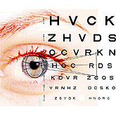
For guidance regarding the conduction of visual acuity, field of vision, heterophoria, and color vision tests, please refer to Items 50-54.
The FAA specifices that the examination of the eyes be directed toward the discovery of diseases or defects that may cause a failure in visual function while flying or discomfort sufficient to interfere with safely performing airman duties.
The Examiner should personally explore the applicant's history by asking questions concerning any changes in vision, unusual visual experiences (halos, scintillations, etc.), sensitivity to light, injuries, surgery, or current use of medication. Does the applicant report inordinate difficulties with eye fatigue or strain? Is there a history of serious eye disease such as glaucoma or other disease commonly associated with secondary eye changes, such as diabetes? (Also see Item 53 and Item 54).
Links to other Portions of the Eye Examination:
Return to Index of Specific Conditions
Return to Part 67 Index
Go Find an AME