
FAA Aeromedical Work UpECHO (Echocardiogram)
|
|
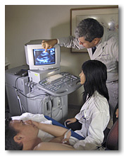

The echocardiogram is an ultrasound of the heart. Thousands of pilots have had this test as part of the recertification process. Using standard ultrasound techniques, two-dimensional slices of the heart can be imaged. The latest ultrasound systems employ 3D real-time imaging.
In addition to creating two-dimensional pictures of the cardiovascular system, the echocardiogram can also produce accurate assessment of the velocity of blood and cardiac tissue at any arbitrary point using Pulsed or Continuous wave Doppler ultrasound. This allows assessment of cardiac valve areas and function, any abnormal communications between the left and right side of the heart, any leaking of blood through the valves (valvular regurgitation), and calculation of the cardiac output as well as the ejection fraction.
Echocardiography is usually performed by cardiologists or cardiac sonographers. Your AME may be experienced in performing and interpreting this test, you he / she may refer you to a cardiologist or other specialist. Regardless of where you get the test, the results will be routed back to your AME and forwarded to the FAA.
Types of Echocardiograms
Transthoracic Echo (TTE)
The standard echocardiogram required by the FAA is also known as a transthoracic echocardiogram, or TTE. In this case, the echocardiography transducer (or probe) is placed on the chest wall (or thorax) of the subject, and images are taken through the chest wall. This is a non-invasive, highly accurate and quick assessment of the overall health of the heart. A cardiologist can quickly assess a patient's heart valves and degree of heart muscle contraction (an indicator of the ejection fraction).
The TTE is commonly used to help diagnose endocarditis. Diagnostic findings by the Echocardiogram include definitive evidence of vegetation or thrombus on valves or other endocardiac structures, abscesses, or disruption of a prosthetic heart valve.
The TTE is highly accurate for identifying vegetations, but the accuracy can be reduced in up to 20% of adults because of obesity, chronic obstructive pulmonary disease, or chest-wall deformities. Transesophageal echocardiography, if available, may be more accurate than TTE because it excludes the variables previously mentioned and allows closer visualization of common sites for vegetations and other abnormalities. Transesophageal echocardiography also affords better visualization of prosthetic heart valves.
TransEsophageal Echo (TEE)
Another way to perform an echocardiogram is to insert a specialised scope containing an echocardiography transducer (TEE probe) into the patient's esophagus, and record pictures from there. This is known as a transesophageal echocardiogram, or TEE. The advantages of TEE over TTE are usually clearer images. The transducer is closer to the heart and doesn't have the ribs and lungs to deflect the ultrasound beam. Some structures are better imaged with the TEE. These structures include the aorta, the pulmonary artery, the valves of the heart, and the left and right atria. While TTE can be performed easily and without pain for the patient, TEE may require light sedation and a local anesthetic lubricant for the esophagus. Unlike the TTE, the TEE is considered an invasive procedure. In some situations, the FAA may require that the airman undergo a TEE instead of a TTE. Work closely with your AME so you know what to expect and avoid unnecessary testing.
In some centers, sedation is used to ease the discomfort to the individual. The use of local anesthetic agents and sedation can decrease the gag reflex, making the ultrasound probe easier to pass into the esophagus. The transducer and cable are then coated in a lubricant, placed in the patient’s mouth, and then passed down the patient's throat. The patient is instructed to swallow while the probe is being passed down, to prevent it from going into the trachea. Although the placement of the thumb-wide transducer is uncomfortable, there are very few complaints of gagging from the patient once the transducer is in the correct location.
Stress Echo
Finally, some of the FAA protocols refer to a "stress echo," which is a specialized form of echocardiogram with greater sensitivity for coronary heart disease. During this test, the pilot has an echocardiogram is done both before and after the heart is stressed either through exercise or by injecting a medicine that makes your heart beat harder and faster. A stress echocardiogram is usually done to find out if you might have decreased blood flow to your heart. Be sure to clarify with your AME which type of Echocardiogram you will need.
Cardiac Problem Index
Overall Problem List
CFR Part 67 Index
Class 1 Standards
Class 2 Standards
Class 3 Standards
Find an AME
Pilot Home
Note: Syncope (fainting), not satisfactorily explained or recurrent requires deferral (even though the syncope episode may be medically explained, an aeromedical certification decision may still be precluded). Syncope may involve cardiovascular, neurological, and psychiatric factors. AMEs will not immediately issue medical certificates if there are heart conditions that require deferral, or for any other cardiac condition that may result in sudden or subtle incapacitation. If the airman has one of these conditions, then the AME will consult with the FAA (AMCD) or the Regional Flight Surgeon. Medical documentation must be submitted for any of these cardiac conditions to support a possible waiver (special issuance) of an airman medical certificate.